Important Facts About Ollier’s Disease
What is Ollier’s disease? Is there a cure? Is it painful? How common is this condition? Here are some important facts to know about Ollier’s disease. This condition is usually not fatal, but can be very painful. This article will answer some of the most common questions about Ollier’s disease, as well as give some information on the different symptoms that you may experience.
Is there a cure for Ollier’s disease?
The diagnosis of Ollier disease is made by specific radiographic findings and clinical manifestations. Histopathological examinations are minimally helpful. This disease usually affects multiple bones, but it can also occur on only one side. Patients usually present with painless bony masses. Plain radiography usually provides sufficient information, but additional testing like ultrasound, magnetic resonance imaging, or scintigraphy is sometimes necessary to confirm the diagnosis.
Ollier disease is a rare non-genetic skeletal disorder characterized by multiple enchondromas in the bones of the limbs. It is often present at birth but may not become apparent until childhood. It usually affects long bones and the cartilage that lines joints. It typically affects the pelvis, ribs, and hands, but can also affect the skull. Treatment for this disorder varies depending on the location, size, and type of enchondromas present.
Currently, no cure is available for Ollier disease, but there are treatments available to control its symptoms. In the meantime, patients should be closely monitored for any changes. Treatment aims at preventing the development of malignant tumors and restoring function.
Is Ollier’s disease painful?
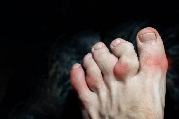
Ollier’s disease is a bone disease that causes cartilage in a joint to forget to turn into bone. Although it can affect people of all ages, most cases are seen in adolescents and young adults. It typically affects the long bones of the legs, but it can also affect the smaller bones of the hands and feet. Symptoms include swelling and lumps in the affected area.
Because of the genetic mutation that causes Ollier’s disease, treatment is mainly focused on stabilizing and strengthening the affected bones. This may include surgery to remove the enchondromas. Other treatments include bone grafts or screws to help the bones heal. Physical therapy may also help with pain and improve mobility.
X-rays may reveal a variety of different types of lesions. This makes it difficult to determine the precise risk of development of a malignant neoplasm. However, it is possible that a patient may develop a tumor in the hip or lower extremity. MRIs, meanwhile, can reveal abnormal bone and soft tissue signals that point to the presence of malignancy.
What is Ollier’s and Maffucci syndrome?
The two diseases Ollier’s and Maffucci are both characterized by a genetic defect known as IDH1. Although neither disease is hereditary, it is a rare and unpredictable disorder that affects both sexes and all ethnic groups. Although the cranium and spine are most frequently affected, the hands and other parts of the body are also at risk. Both disorders are characterized by calcifications of soft tissue and multiple enchondromas.
Patients with Ollier’s disease have abnormal bony development and cartilage enchondromas. The absence of cutaneous vascular lesions makes the disorder difficult to detect until early childhood. Ollier’s disease affects the long bones and joints in the legs and arms. It causes enchondromas to grow from cartilage in the bones, and causes bone shortening. This condition can also affect multiple bones and may lead to scoliosis.
Maffucci syndrome is related to Ollier’s disease and is characterized by multiple enchondromas throughout the skeletal system. In addition to the enchondromas, patients with Maffucci syndrome have abnormal blood vessels under their skin called hemangiomas. These blood vessels may form into tumors that can cause pain. These tumours are often non-familial and appear spontaneously.
How common is Ollier disease?
The prevalence of Ollier’s disease has not been established, but some studies suggest that one in 100,000 people have it. However, this estimate is likely low due to under-reporting of mild cases. The disease is present in both sexes, although men are more likely to suffer from it than women. The disease does not have any obvious symptoms at birth, so it is usually not diagnosed until later in life. However, it can be detected with radiography. Most studies indicate that Ollier’s disease is sporadic, although some cases may be hereditary.
The disease is not a life-threatening condition. However, it can lead to limb deformities. While it is treatable, there is currently no cure. In some cases, surgery may be necessary to correct the condition. There are no known causes for the disease, but genetic factors play a role.
Ollier’s disease can be associated with other disorders, including Maffucci syndrome, which causes multiple enchondromas in skeletal tissue. These tumors may be benign or malignant. Patients with multiple enchondromas have a greater risk of developing chondrosarcoma, a rare but serious form of the disease. In some cases, they may also develop soft-tissue hemangiomas.
What causes Ollier’s disease?
Ollier’s disease is a common disorder that affects the skeletal system. Patients may be born with the disease, or develop symptoms as they grow older. The main symptoms of this disorder are bone deformities and discrepancies in the length of limbs. Those with this disease are also at risk for malignant chondrosarcoma. It most commonly affects the femur and tibia bones. Other bones are less commonly affected, including the spine and metacarpals.
In the majority of cases, Ollier’s disease is asymptomatic. It is often diagnosed as an incidental finding during imaging. Radiography will show a bony mass that is elongated or oval-shaped with a well-defined bone margin. Further imaging may reveal calcifications or granular opacities.
While the exact cause of Ollier disease is unknown, scientists have uncovered mutations in the IDH1 and IDH2 genes. These mutations occur during early development, and are present in many enchondromas and cell lines of the disease. Moreover, the relationship between the mutations and symptoms is not clear. Some patients are also affected by mutations in other genes.
How does osteochondroma affect the body?
Osteochondromas are bony outgrowths of cartilage. They typically form on long bones, such as the pelvis, ribs, or scapula. They can be pedunculated or sessile and range in size. Although the condition can be treated, it is often necessary to have the tumor surgically removed.
Osteochondromas are a rare form of bone tumors, which develop on long bones. Unlike other tumors, they are noncancerous and do not cause symptoms. However, multiple osteochondromas in a person may result in Gardner syndrome, requiring surgery.
Osteochondromas form when a developmental signaling pathway is disrupted. This pathway controls skeletogenesis. Scientists have discovered that mice with two null alleles of the Ext1 gene do not reliably form osteochondromas. However, when mice are heterozygous for both genes, they display stereotypic osteochondromas along long bones.
Osteochondroma is an abnormality of the cartilage that lining the bone is made up of. It develops on the surface of the bone, typically near the growth plate, the area of cartilage that hardens into bone as the child grows. While osteochondroma is generally painless, the patient can seek surgery if it causes pain or threatens to fracture.
Can osteochondroma turn cancerous?
Most osteochondromas are benign and do not cause any symptoms. They can occur sporadically or in clusters. In children, osteochondromas can be diagnosed through an x-ray, which uses a small amount of radiation to see if there is a mass. Usually, osteochondromas show up as a hard mass near the growth plate.
When osteochondroma is suspected, patients will need to have a thorough evaluation. This may include imaging scans, including MRI or CT scans. MRIs are often used to see whether there is cancer present, as they can provide a cross-sectional view. A biopsy of the tumor’s surface is also used to identify any changes. If osteochondroma is not cancerous, treatment will typically include monitoring the tumor over time.
The risk of developing a cancerous osteochondroma is higher in people with multiple osteochondromas. Multiple osteochondromas can occur in males than females, and their number and location can vary from individual to individual. A person with multiple osteochondromas may experience more complications and interfere with the growth of normal bone.
Do osteochondromas need to be removed?
Osteochondromas in Ollivier’s disease are often benign, and may not require treatment. However, if the condition is accompanied by symptoms or if the tumor is causing pressure on nearby bones, blood vessels, or tissues, surgery may be necessary. The risks associated with osteochondroma surgery are minimal, and patients can go home the same day as the surgery.
Osteochondromas are non-cancerous bone tumours that usually begin near the ends of long bones. They are the most common type of benign bone tumour. Most often they develop in childhood, and typically stop growing once the skeleton has stopped growing. However, some people may develop a cancerous osteochondroma.
Osteochondromas are common in children, and usually occur at the knee, pelvis, or proximal femur. These tumors can be painful and cause deformities. However, only about 5% of patients develop malignant osteochondromas.