What is Olliers Disease?
Olliers disease is a limb abnormality that affects children. There is no known cure for Olliers disease. Most cases present in early childhood, and there is no obvious sex-related tendency. Children affected by Olliers disease have limb abnormalities that affect their growth and development.
Is there a cure for Ollier’s disease?
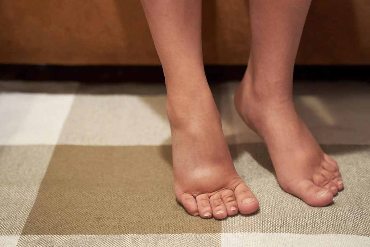
Ollier’s disease is a condition in which the cartilage in the bones fails to develop into bone. It can occur in people of any age, but most cases are seen in young children and young adults. It often affects small bones in the hands and feet. The disease typically causes pain and swelling in the affected area. In some cases, it can lead to a lump.
There is no known cure for Ollier’s disease, and the risk of malignant transformation is unknow. Few studies have been done to quantify this risk, and there is no screening protocol for this condition. Ollier’s disease is named after Louis Xavier Edouard Leopold Ollier, a French surgeon who was born in 1830. He graduated from the Lyon Hospital in 1851 and later became a professor of clinical surgery.
A diagnosis of Ollier disease is often made based on the clinical presentation and specific radiographic findings. Histopathologic examinations may have a limited role in confirming the diagnosis. The disease usually affects two or more bones, but there are some cases in which one bone is affected over the other. Patients usually experience bone masses in early childhood. Ollier’s disease can affect a child’s growth, and an appropriate diagnosis is essential.
Is Ollier’s disease painful?
Ollier’s disease is a rare disorder that affects the joints of the spine and extremities. It is caused by multiple enchondromas, benign growths that occur near the growth plate cartilage. This disorder is often asymptomatic and non-familial. In 8.5 percent of patients, these tumors progress to malignancy. Surgical treatment is usually indicated in early-onset cases.
The pain associated with Ollier’s disease is usually mild and not accompanied by other symptoms. Patients can experience limitations in movement of their skeletal system and a mild wasting of the muscles. However, the disease is not life-threatening, and most patients with Ollier’s disease can lead normal lives. Ollier’s disease is not inherited and occurs when cartilage fails to convert into bone. It is most common on the hands and feet, but can affect any part of the skeleton. Common symptoms include swelling and a lump.
Treatment of Ollier’s disease is usually conservative. Surgical intervention is sometimes needed in severe cases. Treatment varies depending on the size and location of the tumours.
How common is Ollier disease?
Ollier disease is a rare skeletal disorder characterized by multiple cartilaginous tumours. These occur in the diaphyses and medulla of short and long tubular bones, most often in the hands and feet. The disease is rare and does not run in families. It is usually diagnosed during childhood and stabilizes over time. The tumors can be removed surgically if they cause mobility problems or weakening of the bones. In some cases, the tumors can be malignant. Although the disease can be fatal in some cases, in most cases, people can lead a normal life.
The cause of Ollier disease is unknown, but genetic changes have been linked to the disorder. The mutations affect the IDH1 and PTHR1 genes. Ollier disease is also associated with a rare genetic disorder called Maffucci syndrome. The gene mutations are somatic, meaning that they are found in only a certain percentage of cells. As such, the risk of passing the disease to children is approximately the same for both males and females.
What causes Ollier’s disease?
Diagnostic tests for Ollier disease include X-rays and magnetic resonance imaging (MRI) to look for abnormal bone and cartilage growth. Patients may also undergo a biopsy. In severe cases, malignant changes in the bone may be detected using an MRI. There are no known cures for Ollier disease, but treatment is focused on identifying problematic enchondromas. In addition to surgery, treatment may include physical therapy or other supportive services.
Ollier disease is a rare, nonhereditary skeletal disorder characterized by multiple cartilage masses in the bones. The disease usually occurs in the fingers, hands, or feet. The symptoms usually appear during early childhood. In some rare cases, the growths may progress to bone sarcomas.
The symptoms of Ollier disease may be present at birth, but are usually not recognizable until early childhood. People with the condition typically have underdeveloped muscles and short stature. In some cases, the lesions associated with the condition may become cancerous, known as chondrosarcoma. People with Ollier disease have an increased risk of developing liver and ovarian cancer.
How does osteochondroma affect the body?
To diagnose osteochondroma, a doctor will look at your medical history and perform a physical examination. He or she will check for pain or tenderness in the area of the bone. He or she will also check the range of motion of the affected area. If the diagnosis is unsure, the doctor can order imaging tests. Computed tomography (CT) scans use large magnets and radiofrequencies to create detailed images of the body. This type of scan shows the bony growth in detail.
In most cases, osteochondromas do not cause any problems. Surgical removal is only needed when the mass becomes too large and starts to press on nerves or blood vessels. Though rare, there is a slight risk that osteochondromas can develop into malignancies later in life. Symptoms may occur at any time during childhood, but usually disappear by the time the child reaches skeletal maturity.
Osteochondromas develop during the first decade of life and cease to grow when growth plates close during puberty. Depending on their size, they can range from one to ten centimeters in diameter. They may be pedunculated or sessile and may be confined to one or more joints. Typically, the pedunculated form is more common.
Can osteochondroma turn cancerous?
Patients with osteochondroma will undergo a physical examination and imaging test to determine the tumor’s size and shape. These tests can include an x-ray, MRI, and CT scan. A biopsy of the affected area may also be performed to determine whether the disease has spread to nearby organs or tissues. Once the diagnosis is made, treatment consists of careful monitoring over time and regular X-rays.
The cause of osteochondroma is unknown, but genetic abnormalities may play a role in its development. Researchers at the Children’s Hospital of Philadelphia are currently working to understand the causes and treatment options for osteochondroma. Diagnostic imaging can produce detailed images of bone structures, soft tissues, and muscles.
Osteochondromas may be benign, or they may become cancerous. If a tumor causes pain or grows in size, it may need to be surgically removed. If it is benign, osteochondromas are often asymptomatic and do not require treatment. The patient should see their doctor if it becomes painful, especially if it has spread to nearby organs.
Should I worry about osteochondroma?
Osteochondromas are bony masses that originate from the epi-metaphysis in long bones. They are typically benign and increase in size with age. These growths often occur for a prolonged period of time before any symptoms are observed. Osteochondromas may be broad-based or pedunculated. They often have a strong signal on T2-weighted MRI images.
The treatment of osteochondromas depends on the size, location, and extent of symptoms. In young children, surgical removal of the affected bone may be indicated if it is disabling or causes joint deformity. Untreated osteochondromas can cause pain or hemothorax. In some cases, osteochondromas can cause short stature.
Osteochondromas are generally considered to be developmental lesions, but in rare cases, they may also be neoplastic. These are caused by inherited mutations in the EXT1 and EXT2 tumor suppressor genes. In these patients, the mutations affect the differentiation and migration of chondrocytes.