Is Liver Parenchymal Disease Dangerous Or Curable?
If you have liver parenchymal disease, you may be wondering whether it is dangerous or curable. Fortunately, this disease is treatable. In this article, you will learn about symptoms and treatment options. This condition is also often reversible. In some cases, it can lead to cirrhosis.
Is liver parenchymal disease serious?
Liver parenchymal disease is a condition in which the cells in the liver become damaged. This can vary in severity and may be caused by various diseases. Your physician can make the diagnosis by looking at your liver’s appearance and function. A doctor can also order a liver biopsy to confirm the diagnosis and determine the cause of the disease. Patients who are diagnosed with parenchymal disease may need to adjust their diet and lifestyle accordingly.
Liver parenchymal diseases are a common health problem affecting 1.5 billion people worldwide. If left untreated, they can lead to cirrhosis, a potentially fatal condition. Because of this, it is critical to detect liver disease early and monitor it closely during treatment. Liver biopsy is the gold standard of liver tissue assessment. However, it has several limitations, including invasiveness, sampling error, and intra-observer variability.
The liver is capable of processing about 30 ml of pure alcohol per hour. Alcohol that exceeds that limit will be processed by the liver until it reaches its capacity. Treatment of specific liver diseases may include medications, including antibiotics, corticosteroids, and bile acids. Supportive therapy may also include diuretics, antivirals, and nutritional therapy. However, in some cases, liver parenchymal diseases can lead to chronic liver injury and may progress to other liver diseases, such as hepatitis.
Is liver parenchymal disease reversible?
The differential diagnosis of reversible liver parenchymal disease is not as straightforward as it would be with more chronic conditions. Despite the wide range of pathological processes that result in the disease, there are certain clinical criteria that must be met in order to correctly diagnose this condition. In addition, it is crucial to diagnose this condition at an early stage, and to monitor its progression during treatment. Luckily, there are a few diagnostic tests that can help in this regard.
Ultrasound is a noninvasive test for the diagnosis of liver steatosis. Generally, it identifies hepatic steatosis by the degree of echogenicity in the liver parenchyma. This imaging technique is also useful in the detection of hepatic fibrosis. The presence of fatty infiltration may cause decreased visualization of the diaphragm, posterior right lobe, and intrahepatic vessels.
In cirrhotic liver, the ECM content is six times greater than that of normal liver tissue. Early injury results in increased deposition of collagen type III and fibronectin. Chronic injury, on the other hand, increases the deposition of collagen type IV, undulin, elastin, laminin, and Hyaluronan. The concentrations of chondroitin sulphate proteoglycans also increase.
Is liver parenchymal disease cirrhosis?
Liver parenchymal disease is a common problem. This disease is characterized by diffuse damage to the liver. Over a period of time, this damage can progress into fibrosis or hepatocellular carcinoma. Early diagnosis of this disease is important for successful treatment and prevention. Several noninvasive techniques have been developed to evaluate liver parenchyma and fibrosis. These methods use serum biomarkers and conventional imaging modalities.
Cirrhosis is a chronic disease characterized by a progressive decline in hepatic function. This condition affects the liver’s synthesis of clotting factors and excretion of bile. It is caused by a variety of different factors, including toxins, alcohol abuse, and genetic disorders. As a consequence, it is a leading cause of death in many countries. It is the second leading cause of death globally after cancer.
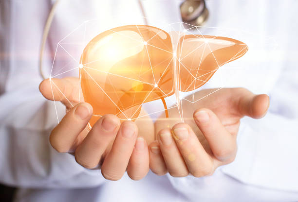
The liver is a large, solid organ that is considered a gland. It is located on the right side of the abdomen. The rib cage protects it. It is divided into two main lobes. The liver cells receive blood from two different sources: the hepatic artery supplies oxygen-rich blood from the heart, and the portal vein delivers nutrients from the intestines and spleen.
How is liver parenchymal disease diagnosed?
There are several ways to diagnose liver parenchymal disease. The most common ones are fatty liver cirrhosis. Histological examination and a multivariate analysis can also help. Depending on the stage, there may be symptoms that don’t seem to be related to the disease.
A biopsy can also be used to diagnose this disease. A biopsy of the liver will provide a tissue sample that can be analyzed for signs of fibrosis or liver failure. Other tests include measurements of liver enzymes and the INR (international normalized ratio). If an INR level is abnormal, it indicates a liver problem. Imaging tests can also detect scarring and tumors. A fibroscan can measure fat deposition and scarring.
Liver cysts can also be detected with ultrasound, computed tomography, or magnetic resonance imaging. These images help the physician diagnose the disease and monitor its growth. Molecular genetic tests can also be performed to detect a genetic mutation in the SEC63 or LRP5 genes. These tests are useful in detecting the disease in individuals who inherited it from a parent. Other blood markers can help diagnose liver disease, including alkaline phosphatase and gamma-glutamyl transpeptidase.
What is the function of liver parenchyma?
The liver plays an important role in many bodily functions, including the production of proteins, blood clotting, and metabolism of glucose and iron. When this vital organ is damaged, illness can result. Liver diseases may be the result of infection, genetic disorder, or toxins, or may develop as a complication of other diseases. When they do, these diseases disrupt the liver’s function and can lead to serious consequences for the body.
The liver parenchyma is traversed by a number of vascular structures. These include the portal vein and hepatic artery. The portal vein supplies oxygenated blood to the liver, while the hepatic artery provides deoxygenated blood. After traversing the portal vein and artery, blood flows through the liver parenchyma and connects to the right atrium.
Liver parenchymal disease involves inflammation of the liver’s parenchyma. This inflammation can result in a variety of outcomes, including hepatocellular necrosis, apoptosis, or fibrosis. The type of inflammation depends on the cause of the inflammation, the level of the lesion, and the response of the body’s immune system. Microscopic examinations can reveal the type of inflammation a person has.
What is normal liver parenchyma?
In the world, approximately 1.5 billion people suffer from chronic liver disease, making it an important global health issue. Several types of liver disease affect the liver parenchyma, causing damage that can progress to hepatocellular carcinoma and fibrosis. An early diagnosis and treatment can greatly influence the course and outcome of these diseases. Fortunately, there are now noninvasive techniques for evaluating liver tissue and parenchyma, including ultrasound and conventional imaging modalities.
Ultrasound can help diagnose hepatic steatosis in adults. However, ultrasound cannot differentiate between fibrosis and normal liver tissue, and it has low sensitivity for detecting cirrhosis. Fortunately, a recent meta-analysis found that US can reliably detect moderate to severe hepatic steatosis in adults. Ultrasound grading varies according to how well the liver parenchyma can be visualized.
What is the parenchyma?
Liver parenchymal disease is an inflammation of the liver. The inflammation results in a variety of cellular changes. Acute hepatitis, for example, is characterized by apoptosis and hepatocellular necrosis. These cellular changes will vary depending on the cause, the host response, and the stage of the lesion. Several different types of inflammation can be detected, including neutrophilic and inflammatory hepatitis.
Liver parenchymal diseases result from many pathophysiological processes. They may progress from early stages of liver injury to more serious forms of liver disease, including cirrhosis. It is important to identify this disease in its early stages and monitor treatment to ensure it is curable. Noninvasive tests, such as ultrasound, are now available to assess liver tissue.
The liver parenchyma is made up of small lobules, called hepatocytes. The lobules contain portal tracts located in the periphery that connect to the central vein. In chronic liver failure, hepatocytes are arranged in cords of cells, which connect the portal tracts. As the liver deteriorates, scar tissue replaces healthy liver tissue. This condition usually progresses over months to years and results in cirrhosis.
What are the two types of parenchyma?
There are two main types of liver parenchymal disease: adenocarcinoma and hepatocellular carcinoma. The former is the most common type, while the latter is rare but can be fatal. While each type requires treatment tailored to the patient’s individual needs, both conditions are often treated through the same surgical procedure.
The liver parenchyma consists of polygonal columnar units called lobules. Each lobule is lined with hepatocytes arranged in a radial pattern around a central vein. Sinusoidal endothelial cells line the spaces between hepatocytes and connect to the central vein.
The first type is chronic cholestatic disease (PBC). It results in the destruction of small bile ducts but does not involve the large bile ducts. The disease is probably autoimmune. It is associated with keratoconjugularitis and Sjogren’s syndrome. It may also be a manifestation of a disorder of the small duct epithelia.


