X-Rays of the Heel Bone and Pathophysiology of Haglund Disease
The X-rays of the heel bone can help your doctor determine if you have Haglund’s deformity. This will also allow the doctor to create customized shoe inserts to help stabilize your foot. If you do have the disease, your doctor will probably recommend an orthotic device. These devices help stabilize your foot while you walk or run.
Surgical treatment options
Surgical treatment options for Haglund disease have improved over the years. Modern techniques utilize smaller portals and allow functional recovery with reduced tissue damage. They may also be combined with endoscopic techniques to achieve minimally invasive surgery. During this procedure, a patient is anesthetized and an incision is made in the heel next to the Achilles tendon. The degenerated tendon and protruding bone are removed and another tendon may be inserted in its place.
Haglund’s deformity is an inherited deformity. Its symptoms can mimic Achilles tendonitis, but a podiatrist can diagnose it by examining the heel. They may also perform MRI or ultrasound to help pinpoint the cause of the condition. Non-surgical treatments for Haglund disease focus on relieving pain and reducing swelling of the bursa.
Surgical treatments for Haglund’s deformity are available for individuals with severe deformity. Both open and endoscopic surgery are effective. Endoscopic surgery is a safe procedure, with lower complication rates and shorter surgical time. However, further research is needed to optimize this technique.
Surgical treatment of Haglund disease includes removing the bumps. Conservative treatments can also be used to reduce the size of the bumps. Conservative treatments focus on less invasive procedures. They can be effective in easing symptoms and improving quality of life. However, some people with the disorder may need other treatment options.
A foot specialist will first examine the appearance and mobility of the foot. Patients with Haglund’s disease usually complain of severe pain in the heel and difficulty walking. The doctor may conduct an X-ray to assess the severity of the problem. An X-ray can reveal a bony protrusion in the heel area. The doctor may also use ultrasound or MRI to evaluate the soft tissues. The X-ray may also reveal calcifications of the Achilles tendon.
Although nonsurgical treatment options for Haglund disease are available, surgical treatment is necessary for patients with severe symptoms. Conservative treatment options may include the use of ice packs to reduce pain and swelling. Physical therapy and soft tissue massage may also be beneficial. Orthotic devices are also available for patients with Haglund disease.
If conservative measures fail to relieve the pain, surgical treatment is the next step. In some cases, the deformity can be removed with minimal damage to the Achilles tendon. Surgery can also involve removing excess bone from the heel to reduce pressure on the Achilles tendon. The patient usually recovers within four to six weeks.
Conservative treatment for Haglund disease consists of lifestyle readjustments and nonsurgical treatment options. Lifestyle changes and the use of correct shoes can reduce discomfort and pain associated with Haglund’s disease. Medications and anti-inflammatory drugs can also help reduce pain and inflammation. But surgery is often necessary for severe cases.
Surgical treatment for Haglund disease entails several risks. The main risks relate to the anaesthetic used and the surgical procedure. The anaesthetist will discuss any risks associated with the anaesthetic.
Pathophysiology
The pathophysiology of Haglund disease is complicated, but there are treatments available. Patients can undergo conservative or surgical treatment to reduce the size of the bony bumps. Conservative treatments include the use of appropriate shoe inserts. If conservative treatment fails, surgical removal of the deformed bone may be required.
Treatment depends on the severity of the condition. Non-surgical treatments such as rest and ice can relieve the pain and swelling in the soft tissues and the bursa. Podiatric doctors, or surgeons who specialize in foot and ankle disorders, may perform surgery to reshape the heel bone. In addition, stretching the Achilles tendon can help prevent the deformity from progressing to full blown Haglund’s disease.
Physical examination can help detect the problem. A lateral X-ray may show the presence of bony protrusions on the heel or calcifications in the Achilles tendon. Ultrasounds and MRIs can also help diagnose the condition. Ultrasound or MRI images of the foot can also show inflammation of the bursa.
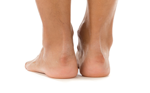
Haglund’s deformity results in a bony growth on the heel. This growth is calcified and irritated, and patients often complain of severe heel pain. The condition also affects the retrocalcaneal bursa, which sits between the Achilles tendon and the heel bone.
Surgical treatment for Haglund’s deformity involves repairing the Achilles tendon and removing the bony lump. The surgeon can perform the procedure through a small incision. The surgeon may also remove some of the bone and tissue. This surgery can provide relief from the pain and reposition the heel bone.
Surgical treatment for Haglund disease is an effective option for pain relief and prevention of further problems. The AAOS recommends surgery for patients with heel pain. Your surgeon will carefully examine your foot and assess the best option for you. The surgeon may file down the protruding bone and Achilles tendon. This will alleviate pressure on the Achilles tendon and other soft tissues in the heel. After surgery, your foot will need up to 8 weeks of rest to heal properly.
Physical examinations are important to confirm the origin of pain and limitations and to determine the anatomical impairment. Physical examinations should be performed in a stepwise process and include muscle strength tests, gait assessments, and special tests. This step-by-step approach ensures that the best possible diagnosis is made.
Outpatient surgery
Haglund’s deformity is usually diagnosed with a physical examination, questions about symptoms, and biomechanical tests. In some cases, X-rays are necessary to confirm a diagnosis of the condition. X-rays can reveal the shape of the heel bone and the alignment of the rear foot joints. They can also show calcification in the Achilles tendon. X-rays also help rule out other possible causes of heel pain. Surgery may be performed to correct Haglund’s deformity.
The procedure is performed through a small incision on the back of the heel. The surgeon then removes the retrocalcaneal bursa and the bony bump causing Haglund’s deformity. After this procedure, the patient may be placed in a splint or surgical shoe to protect the ankle. Patients usually recover fully in four to six weeks.
Patients with this syndrome may also have hereditary heel protrusion. This irritates the soft tissue, causing pain and discomfort. Orthopaedic insoles can be helpful in alleviating the pain in the heel area. Proper fitting shoes are also essential to relieve the pain. Patients should avoid tight shoes, since they can compress the front of the foot.
The procedure is performed under general anesthesia with a local nerve block. The incision is made on the inside of the heel, close to the Achilles tendon. The surgical team then removes the protruding bone and the Achilles tendon. Sometimes, a new tendon is implanted to replace the damaged tendon.
Patients with Haglund’s disease typically report pain in the heel and difficulty walking. This condition is often accompanied by swelling and redness of the inflamed tissue. Surgery may be the only option to relieve pain and restore mobility. During the procedure, a bone spur is removed.
Haglund’s deformity is caused by an overpronated bone at the back of the heel. This bone enlargement can also lead to painful bursitis, an inflammation of the fluid-filled sac between bone and tendon. This enlargement can affect a person’s ability to walk and may affect their life.
During the recovery process, patients are usually able to resume normal activities within a month. In some cases, patients can even return to sports activities after 12 weeks. However, it’s important to check with your doctor before attempting a sports activity. While the recovery process is relatively short, patients should expect some discomfort during the first few months.