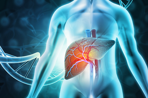
Genetic Liver Disease
The genetic makeup of the liver plays an important role in determining the risk of developing certain forms of liver disease. There are many different types of genetic liver disease, and different genetic mutations are associated with different degrees of liver disease. Genetic testing for these conditions is limited, due to the number of possible mutations and the varied clinical expression of these conditions. Nevertheless, screening tests for genetic liver diseases are an important part of routine care and may help prevent end-organ damage.
Golgi homeostasis
Golgi homeostasis is a central sorting hub in the liver that sorts cargo molecules and delivers them to the appropriate cellular locations. It is a flattened ribbon-like structure that coordinates glycoprotein trafficking and plays an important role in liver function.
A recent study shows that RL disrupts ER-Golgi trafficking by disrupting the ATF6 branch. It also decreased colocalization between the Golgi and ER under the influence of RL and specific ER stress-inducing agents. Golgi fragmentation was found to be triggered in RL-treated cells, but it was not induced in cells treated with TG or RTV.
Golgi structure is subject to remodeling in response to cytotoxic agents and stress. Alcohol, a known hepatotoxin, has been shown to alter Golgi structure in hepatocytes. In addition, anti-Golgi antibodies have been detected in sera of patients with end-stage liver disease. These antibodies are specific to giantin, the highest-molecular-weight protein matrix protein.
Giantin, Rab6a, and NMIIB are essential for Golgi biogenesis. However, they compete for the Rab6a GTPase and de-dimerization of these proteins causes alterations in the Golgi structure. Once EtOH treatment is completed, the Golgi regains its compact structure. Moreover, knockdowns of Giantin, a protein that regulates the transport of selected hepatic proteins, impairs recovery in EtOH-treated cells.
The study of MFSD1 has identified a new transporter in the Golgi. It co-localizes with LAMP-proteins and lysosomes. However, it does not rescue MFSD1 from the lysosomes.
Dubin-Johnson
The genetic code for Dubin-Johnson syndrome (DJS) is unknown. It has an autosomal recessive inheritance pattern. In cases of DJS, a yellow waste substance accumulates in the liver. Symptoms include yellow skin, whites of the eyes, and a discolored tongue. This yellowness occurs indoors and outdoors. Jaundice usually appears on the face first, but it can also affect the chest and stomach.
Patients with DJS have a decreased capacity to metabolize bilirubin. Bilirubin excretion from the liver is affected, and conjugated bilirubinemia is a common complication. The condition is also associated with a decreased ability to excrete bilirubin from the bile canaliculi. This condition affects both males and females equally. It also affects patients who are pregnant.
Dubin-Johnson syndrome is a rare genetic disease affecting the liver. A mutation in a gene involved in the production of bilirubin causes the liver to fail to release the yellow substance into the bile. This accumulation clogs up the biliary system, causing symptoms such as jaundice.
In a recent study, researchers found that a gene called ABCC2 causes Dubin-Johnson syndrome. Mutations in this gene result in predominantly conjugated hyperbilirubinemia. There have been many studies conducted worldwide on this gene, but the genetic pattern of this disease in China has not been determined. The aim of the present study was to investigate this gene’s mutation pattern in Chinese patients. This was done through Sanger sequencing, a method that allows for the identification of multiple mutations in a single DNA segment.
Rotor syndrome
Rotor syndrome is a genetic liver disease characterized by a buildup of unconjugated and conjugated bilirubin in the blood. In this disease, the liver is unable to process the bilirubin efficiently, resulting in jaundice. Rotor syndrome is rare and does not require treatment. Like Dubin-Johnson syndrome, it is not life threatening.
Patients with Rotor syndrome typically have asymptomatic liver disease. However, some may develop generalized non-pruritic jaundice during their childhood. Rotor syndrome patients also experience fatigue and dark urine. They may also develop gastric abnormalities and fever. Genetic testing can help identify this disease.
The disease is inherited in an autosomal recessive pattern, involving mutations in both copies of the SLCO1B3 gene in each cell. This means that patients with Rotor syndrome must have mutations in both SLCO1B1 and SLCO1B3 genes in order to develop the condition. The parents of Rotor syndrome patients carry the altered copies of both genes, but do not show symptoms.
Rotor syndrome is a rare genetic liver disease caused by inactivation of two proteins in the liver. These proteins are responsible for reabsorption of conjugated bilirubin. A defect in either one of these two proteins causes increased bilirubin levels and porphyrinuria. In our case, both genes were inactivated, so the condition was categorized as a form of RS.
Alpha1-antitrypsin deficiency
Alpha1-antitrypsin deficient (A1ATD) is one of the leading causes of genetic liver disease in children. It is a protein-folding disorder characterized by the accumulation of toxic insoluble ATZ proteins in hepatocytes. This accumulation results in inflammation, fibrosis, and cirrhosis. Patients also have an increased risk of hepatocellular carcinoma. Patients with A1AT-deficient livers often require orthotopic liver transplantation, which is curative. However, small-molecule therapies are emerging as potential therapeutics for preserving the native liver and preventing hepatotoxicity.
Alpha-1 antitrypsin deficiency is an inherited disorder that affects the liver and lungs. Symptoms of this disorder may range from jaundice in newborns to poor feeding in adults. Alpha-1 deficiency may also lead to lung disease, including emphysema and panniculitis. People with this disorder may also have other problems, such as fatigue and poor appetite.
AAT deficiency is often caused by an aberrantly folded AAT protein. The most common AAT deficiency variant is the Z allele, which results in an inefficient folding of the protein. The wild-type AAT protein is secreted from the hepatocytes, but the Z mutant protein folds incorrectly and never reaches the trafficking and secretion pathway. In homozygous patients, AAT levels in the serum are reduced by about 10% to 15% compared to normal WT patients.
In patients without alpha1-antitrypsin deficency, the diagnosis is made at an early age of thirty or forty, although there are no other genetic deficiencies that are as closely related to emphysema. Pediatricians who encounter a newborn with jaundice or bleeding are prone to consider more complicated diseases. Fortunately, there is a simple blood test for this disorder.
BSEP
There is a correlation between BSEP and genetic liver disease. A genetic mutation in BSEP causes an abnormally short or inactive BSEP protein, which can lead to severe liver disease. People with this genetic disorder may develop hepatic encephalopathy or hepatocellular carcinoma.
The ABCB11 gene is responsible for human BSEP and contains 28 exons. It is located on chromosome 2’s long arm. This gene has variants that suggest that it is involved in the development of ATD-induced liver damage. Additionally, administration of INH/RMP significantly decreased BSEP expression in mouse livers.
BSEP plays a physiologic role in the secretion of bile salts from the liver. It has been associated with several forms of cholestasis, including familial cholestasis type 2 and acquired cholestasis. More than 100 BSEP mutations have been identified worldwide in independent and collaborative studies. Some of these mutations are missense, while others are nonsense mutations.
BSEP is an important gene involved in many liver diseases. Previous studies have identified a common BSEP polymorphism (V44A) as a genetic risk factor for DILI in Caucasian patients. However, the polymorphism has not been found to be a significant risk factor for the development of DILI in Chinese patients. These previous studies also tended to focus on drug and ethnicity-specific causes.
Genetic abnormalities in BSEP are associated with end-stage liver disease. Genetic alterations in BSEP can cause liver failure, hepatocellular carcinoma, and cholestasis. Moreover, post-translational regulation of BSEP affects intracellular trafficking and functional expression in the liver. Additionally, BSEP expression on the canalicular membrane is altered by ubiquitin-proteasome mediated degradation.
MDR3
To determine whether MDR3 genetic variation in PBC patients reflects the disease, researchers have compared sequence diversity in PBC patients with healthy controls. They found that both groups had similar haplotypes. In addition, the investigators found no significant differences in the number of population-specific variants. However, the researchers did find differences in the number of disease-causing variants.
MDR3 mutations are associated with cholestasis in pregnancy. They can also cause cholestatic liver disease in children. Affected individuals may also experience biliary cirrhosis, jaundice, or scarring of the liver. Mutations in the ABCB4 gene cause MDR3 deficiency. In most patients, the disease follows an autosomal recessive inheritance pattern. However, autosomal dominant inheritance may also occur.
MDR3 genetic liver disease is caused by a mutation in the ABCB4 gene on chromosome 7q21.1. This protein functions in the liver, and deficiency results in non-micellar bile acids accumulating in the hepatocytes. In people with MDR3 deficiency, the disease may progress to end-stage cirrhosis in the fourth decade of life.
The MDR3 gene regulates canalicular phospholipid transport in hepatocytes. Mutations in MDR3 result in a subtype of familial intrahepatic cholestasis. In addition to causing liver failure, MDR3 mutations may also cause biliary cirrhosis. The frequency of MDR3 mutations is unknown. There have been no Asian patients with MDR3 mutations.