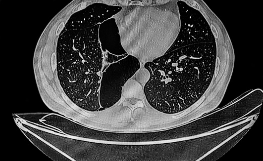
The incidence of incidental bullous lung disease on CT imaging is increasing. This complication is often found in patients after a readmission for spontaneous pneumothorax or COVID-19 pneumonia. This condition is not fatal. However, it is important to identify and treat this disease in the earliest stages.
What causes bullous in lungs?
Bullous in the lungs is a condition that affects the air sacs of the lung. It is often seen on x-rays and is a sharp downward indentation that occurs between the hemi-diaphragm and the adjacent chest wall. Patients may experience pain or discomfort when they speak and are easily treatable.
Several different types of bullae can be found in the lungs. One is known as idiopathic giant emphysematous bulla, which occurs in the upper lobes of the lung. It is most common in young smokers. If the bullae are inflamed, they can interfere with lung function, and rupture may result in vanishing lung syndrome. The condition can be treated and diagnosed with an appropriate medical evaluation.
Another type of bullae is known as a pneumatocele. These apical cysts are one centimeter in diameter. They can occur in a variety of conditions, including bullous emphysema. A patient with bullous emphysema is at increased risk for developing lung cancer. The incidence of lung cancer associated with bullous emphysema is 6.1%, or 3.6 times higher than that of patients without bullous emphysema. There are three possible explanations for the increased incidence of lung cancer among patients with bullous emphysemas. The bullous wall may contain an underlying condition that is causing the material to accumulate. Alternatively, the cancer may arise from a scar in the bullous wall.
How do you get rid of Bulla in your lungs?
The first step in treating bullae in the lungs is to stop smoking. Smokers are at risk for developing the disease as their bullae are large and may cause problems for them. Surgical treatment may be necessary in cases where the bullae are large and blocking lung function.
If left untreated, bullae can lead to lung collapse. Patients with COPD or emphysema are at risk for developing bullae. People exposed to gas fumes may also be susceptible. There are a number of non-surgical treatments for bullae. These include supplemental oxygen and bronchodilators. In addition, pulmonary rehabilitation can help improve symptoms.
Treatment of bullae in the lungs varies depending on the size and location of the bulla. Most commonly, doctors use a combination of medication and surgery to treat COPD. The goal of BE treatment is to reduce or eliminate the risk of the disease.
Can bullous lung disease be cured?
The rate of progression of bullous lung disease varies depending on the severity of the disease and other factors. Early intervention, especially smoking cessation, is the most important step in the treatment of bullous emphysema. The bullae of affected patients often shrink due to an inflammatory response and may even disappear.
Surgical resection of the bulla may help to alleviate symptoms. This is a good option for patients with severe disease, particularly when the bullae are large and compress adjacent lung tissue. The procedure may be performed only after a pulmonary function test shows that the bulla is not contributing to functional gas exchange.
After the resection, patients may receive further therapy. The goal of treatment for bullous emphysema is to improve the patient’s quality of life and lung function. Some treatments, such as steroid inhalers, can help patients overcome shortness of breath and other symptoms. In addition to these, some patients may require antibiotics.
Can bullous emphysema be cured?
Bullous emphysema is a condition characterized by enlarged airspaces or bullae in the lungs. They are usually demarcated by hairline shadows. They are a secondary complication of emphysema. Surgical treatment can reduce the volume of the affected lung.
There are many surgical procedures available to treat this condition. These procedures may help to improve lung function and quality of life in some patients. The removal of large bullae may be effective for some patients. Some patients may benefit from lung transplantation. It is important to note that surgery is not a cure for bullous emphysema.
Smoking is one of the main causes of bullous emphysem. Genetic factors, such as alpha-1 antitrypsin deficiency, may also be a cause of the disease. In addition, intravenous drug abuse may lead to inflammatory damage in the alveoli. Approximately 120,000 people die from bullous emphysemia every year.
Can lung bullae be cancerous?
Bullae on the lungs are often benign, but they can be a sign of more serious disease. They appear as dark, sharply-pointed indentations in the lung parenchyma. If they’re open, they may become infected. If they’re large, however, they can indicate a potentially malignant condition.
The authors of the study found that five of the six patients were diagnosed with lung cancer within a month of the initial hemoptysis. Their initial CT images were so complicated that the cancer was not suspected. Ultimately, they had to perform resections to identify the cancerous lesions. The authors also found that there was fluid collection within the bullae, which made it difficult to determine the wall thickness or mass.
Although the frequency of bullous lung disease is unknown, it does closely follow the epidemiology of COPD. It is more common in older people and those with significant exposure to tobacco smoke.
Can you feel a lung bulla?
A lung bulla is a lump that is about the same diameter as the lung itself. It may be infected or non-infected. If you suspect that you have a bulla, it is important to consult a physician. Bullas are not as dangerous as lung abscesses, but they can still cause serious complications.
Bullas are not cancerous, but they can cause a pneumothorax – a collapsed lung. This is a potentially fatal disease. It can be a sign of an infection or a complication such as a respiratory tract infection. It can also be a warning sign of a bigger problem, such as an underlying heart condition.
Often, patients with COPD will develop a bulla. One case report described a young patient with bronchial asthma who developed a giant lower lobe bulla.
How do you know if you have bullae?
Bullae are large, fluid-filled blisters that can occur on your skin. They’re often a sign of an underlying health problem. Bullae can be painful and can require medical attention. They usually measure more than 5 millimeters in diameter. Some forms of bullae can be prevented or treated. Learn what to look for and how to treat them. Bullae are a sign of a bacterial or viral infection.
If you have bullae, consult a dermatologist. The area should be covered to prevent infection. You can also apply bandages to protect the area. You may also need to have your bullae checked by a physician if you have any bleeding or redness. A doctor can also prescribe antibiotics for an infection that’s affecting your skin.
If your CT scan shows a bulla in the thorax, it could be indicative of another underlying condition. For example, if you have a massive bulla, it could be indicative of a disease like Ehlers-Danlos or Marfans. These conditions are caused by abnormalities in connective tissue, and the presence of these abnormalities will raise the risk for a pneumothorax.